ABOVE: © ISTOCK.COM, Kseniia Glazkova
Most humans move a lot less than our ancestors did, a change in use which is likely reflected in the composition of muscles themselves. Understanding our muscle makeup is a key step towards improving regenerative medicine for neuromuscular diseases. But it has been difficult to quantify aspects of how muscles have changed, including fundamental details such as what fibers exist in which parts of the muscle. Now, a group of scientists has found a way to better identify these fibers. Using an integrated mass spectrometry method, the scientists discovered a group of “superfast” muscles in mice, they reported February 3 in Science Advances.
Muscles are made up of slow twitch fibers, which produce smaller muscle contractions and are more fatigue resistant, and two types of fast twitch fibers, which produce bigger contractions in shorter spurts. However, studies have suggested that repeated exercise can actually cause muscle fibers to switch types, and scientists have even speculated that if they used the right technology, they might find intermediate types that blur the lines between slow and fast twitch fibers. Ng Shyh-Chang, a stem cell biologist at the Institute of Zoology at the Chinese Academy of Sciences and the Beijing Institute for Stem Cell and Regenerative Medicine who coauthored the new study, tells The Scientist that he wanted to examine these fibers more closely and identify which metabolic subtypes exist in which part of a muscle.
The most ideal method for researching this would be to use mass spectrometry, Ng says. He explains that the tried-and-true mass spectrometry protocol, called liquid chromatography mass spectrometry (LC-MS), involves crushing tissue into a semi-liquid mixture called a slurry, then running that the metabolites from that fluid through specialized chromatography columns followed by a mass spectrometer to separate and identify important proteins and metabolites. Although the LC-MS method produces strong signals, which make it easy to identify which basic fiber types are in the sample, Ng says that because the method uses crushed up rather than intact tissue, it is difficult to figure out how exactly those fibers are distributed, and identify rare fiber subtypes.
To overcome this issue, Ng and colleagues decided to examine the fiber distribution by using mass spectrometry imaging (MSI), which can capture high resolution images of intact frozen muscle sections. The type of MSI Ng aimed to use, a process called matrix-assisted laser desorption (MALDI) MSI, involves treating frozen muscle sections with a matrix material, then hitting them with a pulsing laser micrometer by micrometer to produce a map of ions that can be traced back to specific cells or fibers of origin. Because MSI can work on intact tissue, it also gives scientists a better idea of which parts of a tissue go where, and how different fibers group together. Although MSI is powerful, Ng says that it can struggle to produce the same strong signals as LC-MS, which can make it hard to figure out what you’re looking at. University of Florida chemist Richard Yost tells The Scientist in an email that “MSI is not at its best when looking for untargeted compounds, particularly at low concentrations. That’s where [LC-MS] shines.”
See “Scientists Use Centrifuge to Discover a Hormone”
Ng and colleagues were faced with a conundrum: They could either get the metabolite resolution of LC-MS or the spatial resolution of MSI. Or, Ng realized, they could simply do both. To increase their chances of success, and in hopes of learning more about fiber subtypes, Ng and his team started with mouse leg muscle tissue, as its structure is relatively simple and its big fibers would produce strong signals, making them easier to identify. Ng decided to run the fibers through LC-MS and process them through existing metabolome databases to pick out signals of different fiber groups. They then used the same fibers to cross-check the LC-MS data with MSI data, helping them link the patterns they saw in the LC-MS with the high resolution MSI images. Using the muscle fibers and LC-MS to train the MSI software to identify metabolic patterns, Ng and his team ended up with an optimized system that combined MSI’s high specificity and level of detail with LC-MS’ ability to pinpoint metabolites at an individual fiber scale.
“The integration of [LC-MS] and MSI makes studies like this far more likely to provide valuable insights,” writes Yost.

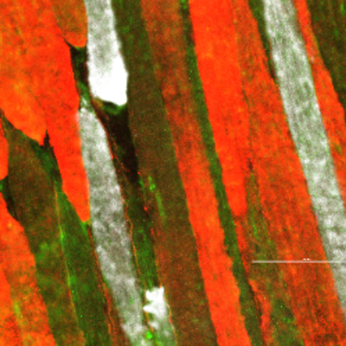
Thrilled that they had gotten the technology working, Ng and his colleagues sifted through the images in hopes of finding something interesting. “We were looking for metabolites, and certain things stood out where we were like, ‘huh, that’s weird, we didn’t expect to see this,’” Ng says. Among the surprises was the pattern of muscle fiber groupings. Going into the experiment, Ng and his team had been looking for the suspected intermediate muscle fibers. Instead, they found that each of the subsets of muscle fibers was very distinct from the other and fell into the categories that had already been identified—save for one exception.
Running a genetic analysis on the fiber that didn’t fit into an existing category, Ng was shocked to find signatures of what are known as “superfast” muscle fibers in the limb muscles of a mouse. These fibers, typically found in areas known for their ultra-high speed and endurance, such as human eyes and hummingbird wings, had never been documented in a mammalian limb bone before. The superfast fibers they found in the mouse limb were slightly different from the ones seen elsewhere in the body, Ng says. Although clinical uses are a long way off, he says that finding these fibers in limb muscles is particularly exciting for regenerative medicine, speculating that because of their fast movement speed, they could be employed to increase neurostimulation and improve neuromuscular coordination in humans.
Knowing that muscle fibers can change types, he says it’s plausible scientists could find a way to artificially induce the production of superfast fibers in the muscles of Parkinson’s patients, but they need to do more research on how these fibers change naturally in the body first.
“Before you saw the fossil dinosaur, you didn’t think that a dinosaur would exist,” he says. “But now that you’ve seen a dinosaur, you can start looking for [more] dinosaurs. So knowing what [superfast muscle] looks like in the limbs allows us to now recognize it and then start looking for factors that can now induce it.”


