What are organoids?
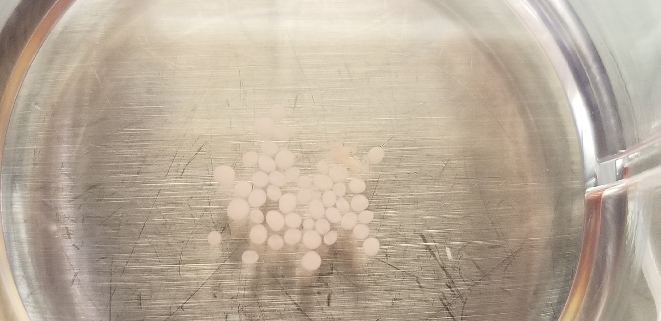
Organoids are 3-D cell cultures derived from pluripotent stem cells that mimic the structure, function, and cellular complexity of human organs. These in vitro, miniaturized versions of organs are especially well suited for studying complex multicellular organ structures, such as the brain, retina, kidney, and lungs, and are now widely used to study organ development and disease.1
Organoids versus Spheroids
Researchers often use common 3-D culture techniques to produce spheroids—round cell clusters of primary or immortalized cells that are popularly used in tumor research.2 Organoids are similar to these structures, except their formation begins with tissue-specific stem cells that self-assemble into microscopic versions of a functioning organ component.1
How Are Organoids Made?
Organoids allow researchers to study matrix-adhered cells and learn about organ development. A process that could take years using live model organisms now takes months in organoid culture. Combined with CRISPR genome editing technology, researchers also use organoid cultures to model genetic diseases in tissues that are otherwise unobtainable, such as the brain. Researchers can produce numerous organoids from tumors or patient-derived induced-pluripotent stem cells (iPSCs), making them ideal models for drug screening and personalized cancer therapies. Organoid culture protocols are continuously improving, giving researchers a chance to focus on novel and targeted treatments while avoiding the risks posed to human subjects.
Organoids are derived from either the directed differentiation of human pluripotent stem cells (hPSCs), tissue-specific adult stem cells, or iPSCs. Because organoids come from active stem cell populations, researchers can expand their cultures repeatedly over time. To create an organoid, scientists embed pluripotent cells in an extracellular matrix such as Matrigel, which serves to support the cells. Specific growth factors and proteins that mimic the in vivo environment maintain the stem cell phenotype. Based on the initial stem cell population and growth factors chosen for a study, the matrix-embedded cells will self-assemble into 3-D organoid structures that behave similarly to a specific tissue.2,3,4

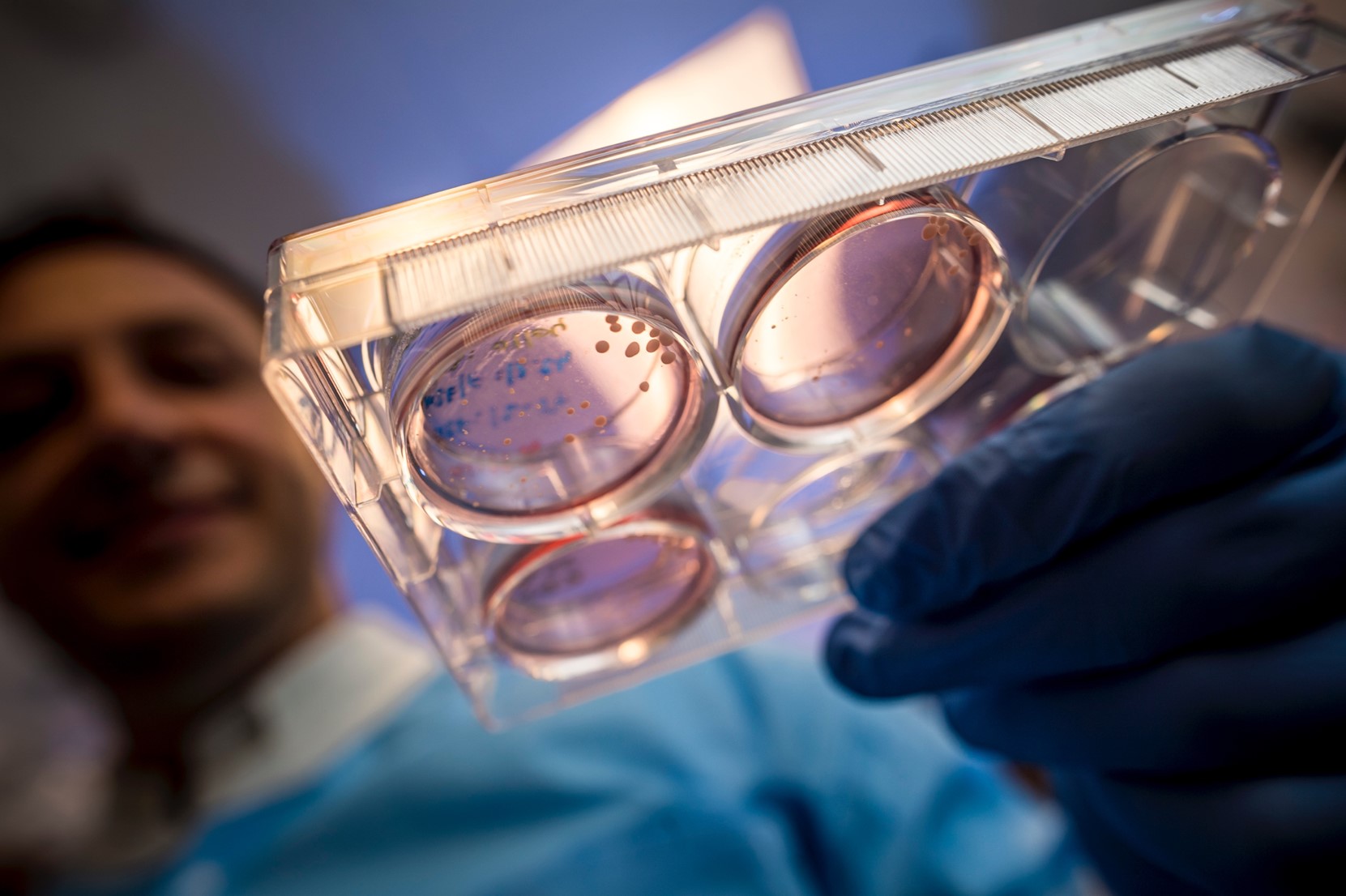
Researchers can adapt most protocols using a typical tissue culture room and standard equipment. When generating organoid cultures with hPSCs, pluripotent cells are initially cultured with a feeder cell population that provides the growth factors needed to maintain stem cell pluripotency. The hPSCs are allowed to form colonies in multi-well plates before they are enzymatically detached from the wells and feeder population. Researchers then dissociate and plate the pluripotent colonies on low-attachment 96 well plates. Over 1-2 weeks, the cells will begin to form embryoid bodies and can be induced towards certain lineages using tissue-specific induction media. Scientists can embed the differentiated embryoid bodies in Matrigel droplets and either cryopreserve or continuously culture them for several months using tissue-specific media.5 There are now also a number of commercial resources that provide pre-generated cryopreserved organoids for purchase.
Types of Organoids and Their Applications
Brain organoids
The brain is a highly complex organ with elements that are unique to humans, so using animal models to study brain development is rather ineffective. Scientists use mainly hPSCs or mouse embryonic stem cells along with neural stimulatory factors in 3-D cell culture protocols. Researchers have designed these protocols to promote cell differentiation into organoids representing specific brain regions. As these brain organoids develop and form multiple tissue layers, they can represent the actual progression of the human brain during gestation. Researchers are continuously improving their methods, pushing the development of these 3-D brain regions further in gestational age to discover unknown brain development mechanisms. For many regions of the brain, however, inducing in vitro development past early gestational ages remains a challenge. Nevertheless, brain organoids provide significant insights into neurodevelopmental, psychiatric, and genetic diseases as well as neural tumors.6
Lung organoids
The lung is another complex tissue that is difficult to study. With its many cell types, highly vascularized structure, and function in oxygen exchange, 2-D in vitro experiments are limiting and in vivo studies are costly. Lung organoids representing alveolar tissues were first developed in 1980, but were not truly successful until 30 years later when Darrell Kotton from Boston University purified lung precursor cells from mouse embryonic fibroblasts and stimulated BMP/FGF signaling to induce lung cell differentiation.7 Techniques have since improved as researchers use hPSCs and iPSCs to model airway epithelial cell function, track early lung bud development, and perform lung cancer screens. The lung is also unique in its ability to regenerate following damage. Therefore, researchers can derive lung organoids from adult stem cells.8 This makes it possible to generate lung organoids from adult patients with genetic or environmental diseases. More recently, lung organoids have become an invaluable tool for studying SARS-CoV-2’s effect on respiratory function and development.9
Gastric/intestinal organoids
The gastrointestinal tract has many functions and consists of multiple germ layers that must work together. In 2011 and 2014, researchers used hPSCs to develop the first intestinal and gastric organoids that contained multiple tissue types.10,11 However, the gastric system is closely tied to neural and immune cells, which made many protocols relatively limited in their ability to recapitulate what goes on inside the body. To make things more complicated, the stomach and intestines have their own microbiomes, which influence multiple processes ranging from the immune response to pharmacokinetics. With this in mind, researchers recently developed protocols that co-culture intestinal commensal bacteria with organoids, which can model gastric diseases and allow researchers to study microbes’ influence on cancer cells.12,13
Cancer organoids
Patient-derived cancer organoids are an invaluable tool for studying cancer progress as well as the design and implementation of precision therapy. Cancer organoid models accurately model patient tumors and scientists use them for basic research and drug screens. Organoids developed from tumors also have a surprising ability to maintain genetic and molecular signatures even when extensively passaged.14 Recently, researchers developed methods to generate and screen patient-derived organoids to determine patient-specific therapies within a matter of days.15
3-D cell culture and assembloids
Researchers have now gone a step further in organ modeling by developing cultured assembloids, which are the next generation of organoids that combine multiple tissues. In organs such as the brain where several different regions and tissue types interact, researchers developed protocols that combine multiple tissues in culture, allowing them to observe and model the tissue interactions that affect cellular function.16 Recently, laboratories adapted these protocols to additional tissues that are increasingly complex, quickly making assembloids an important tool in modeling disease.
References
- M.A. Lancaster, J.A. Knoblich, “Organogenesis in a dish: modeling development and disease using organoid technologies,” Science, 345(6194):1247125, 2014.
- F. Hirschhaeuser et al., “Multicellular tumor spheroids: an underestimated tool is catching up again,” J Biotechnol, 148(1):3-15, 2010.
- M. Huch, B.K. Koo, “Modeling mouse and human development using organoid cultures,” Development, 142(18):3113-25, 2015.
- K. Kretzschmar, H. Clevers, “Organoids: modeling development and the stem cell niche in a dish,” Dev Cell, 38(6):590-600, 2016.
- M.A. Lancaster, J.A. Knoblich, “Generation of cerebral organoids from human pluripotent stem cells,” Nat Protoc, 9(10):2329-40, 2014.
- J. Sidhaye, J.A. Knoblich, “Brain organoids: an ensemble of bioassays to investigate human neurodevelopment and disease,” Cell Death Differ, 28(1):52-67, 2021.


